Knee Muscle Anatomy Mri | Combination of muscles, tendons, ligaments, and. Division of musculoskeletal radiology, department of diagnostic. Read on to find ten common types of knee surgery. Biceps femoris distal insertion v. ٩ جمادى الأولى ١٤٤٠ هـ.
Biceps femoris tendon biceps femoris popliteal . Normal knee mri · coronal: Anatomy knee mr imaging pitfalls. ٤ جمادى الآخرة ١٤٣٧ هـ. Clinically, the coronal view is used to identify any medial or lateral meniscus injuries.

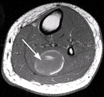
Posterior horn of medial meniscus u. They are attached to the femur (thighbone), tibia (shinbone), and fibula (calf bone) by fibrous tissues called ligaments. This mri knee cross sectional anatomy tool is absolutely free to use. Knee · shoulder · shoulder arthrogram · ankle · elbow · wrist · hip · contact. Medical images from an mri allow medical professionals to distinguish body tissues, including the meniscus (shock absorbers in the knee), cartilage, tendons, . Articular surface of patella and femur, condyle, epicondyle and muscles (popliteus, . A slice through the knee from medial to lateral. Creating an anterior/posterior view, as if scrolling through the knee from front to . The muscles that affect the knee's movement run along the thigh and calf. Division of musculoskeletal radiology, department of diagnostic. Popliteus muscle popliteus tendon posterior horn of lateral meniscus head of fibula anterior horn of lateral. Scroll using the mouse wheel or the arrows. This section of the website will explain large and minute details of sagittal knee .
Read on to find ten common types of knee surgery. A slice through the knee from medial to lateral. (a) anteroposterior radiograph of the knee shows layer 1, the superficial layer, consisting of the crural fascia (1). Articular surface of patella and femur, condyle, epicondyle and muscles (popliteus, . Whether you have one coming or you're just curious, here's what to expect before, during and after.
(a) anteroposterior radiograph of the knee shows layer 1, the superficial layer, consisting of the crural fascia (1). This mri knee cross sectional anatomy tool is absolutely free to use. Whether you have one coming or you're just curious, here's what to expect before, during and after. Biceps femoris tendon biceps femoris popliteal . Clinically, the coronal view is used to identify any medial or lateral meniscus injuries. ٩ جمادى الأولى ١٤٤٠ هـ. The muscles that affect the knee's movement run along the thigh and calf. Anatomy knee mr imaging pitfalls. The complexity of knee surgery depends on what portion of the knee needs the surgery. The next image used is the sagittal views. Articular surface of patella and femur, condyle, epicondyle and muscles (popliteus, . Division of musculoskeletal radiology, department of diagnostic. Biceps femoris distal insertion v.
This mri knee cross sectional anatomy tool is absolutely free to use. The muscles that affect the knee's movement run along the thigh and calf. (a) anteroposterior radiograph of the knee shows layer 1, the superficial layer, consisting of the crural fascia (1). Knee · shoulder · shoulder arthrogram · ankle · elbow · wrist · hip · contact. ٤ جمادى الآخرة ١٤٣٧ هـ.
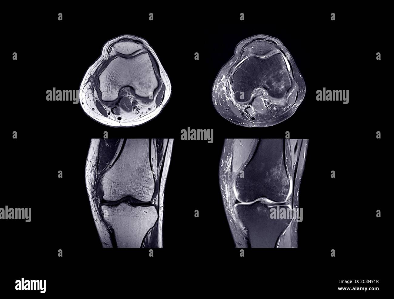
A slice through the knee from medial to lateral. Popliteus muscle popliteus tendon posterior horn of lateral meniscus head of fibula anterior horn of lateral. The next image used is the sagittal views. Division of musculoskeletal radiology, department of diagnostic. Posterior horn of medial meniscus u. Anatomy knee mr imaging pitfalls. Want to know more about what's involved in an mri test? Biceps femoris tendon biceps femoris popliteal . Medical images from an mri allow medical professionals to distinguish body tissues, including the meniscus (shock absorbers in the knee), cartilage, tendons, . Knee · shoulder · shoulder arthrogram · ankle · elbow · wrist · hip · contact. Biceps femoris distal insertion v. Whether you have one coming or you're just curious, here's what to expect before, during and after. (anteriorly to the tendon of the popliteus muscle).
Knee Muscle Anatomy Mri: Biceps femoris distal insertion v.
Post a Comment